|
|
|
|
BIOLOGICAL EVIDENCE FOR CRIME INVESTIGATION
Hariom Rajput 1![]()
![]()
1 National
Crime Bureau of Investigation, Ministry of Health and Family Welfare,
Government of India, India
|
|
|
ABSTRACT |
|
|
The review paper helpful for knowledge and current data biological evidence in forensic science is pivotal to maintaining the integrity of biological evidence and ensuring the reliability of forensic analyses. This encompasses a set of strategies aimed at preventing contamination during evidence collection, handling, and analysis. Key elements include the use of Personal Protective Equipment (PPE), such as gloves, masks, and protective clothing, to minimize direct contact and contamination from the environment. Sterile tools and evidence immediately after collection. Environmental controls are also critical. Workspaces must be kept clean, and access to crime scenes and evidence storage areas should be strictly controlled. Airflow management, such as the use of laminar flow hoods, helps to reduce airborne contaminants. Detailed documentation and maintaining a clear chain of custody are fundamental to ensure that each piece of evidence can be traced and accounted for, reducing the risk of tampering or unauthorized access. Training and dedicated equipment for DNA analysis and the implementation of controls during DNA extraction and amplification processes to detect any contamination. Laboratory practices should include separate areas for different stages of evidence processing and regular decontamination of work surfaces and equipment. By strictly following these protocols, forensic laboratories can significantly reduce the risk of contamination, thereby ensuring the credibility and accuracy of their findings in the justice system. |
|||
|
Received 28 July 2024 Accepted 02 August 2024 Published 18 August 2024 Corresponding Author Hariom
Rajput, hariomraj9171494082@gmail.com
DOI 10.29121/DigiSecForensics.v1.i1.2024.23 Funding: This research
received no specific grant from any funding agency in the public, commercial,
or not-for-profit sectors. Copyright: © 2024 The
Author(s). This work is licensed under a Creative Commons
Attribution 4.0 International License. With the
license CC-BY, authors retain the copyright, allowing anyone to download,
reuse, re-print, modify, distribute, and/or copy their contribution. The work
must be properly attributed to its author.
|
|||
|
Keywords: Biological Evidence, Investigation, Buccal Smear,
Blood Smears, DNA, Analysis, PCR, Techniques, Forensic |
|||
1. INTRODUCTION
Biological investigations are essential for understanding the intricate mechanisms of life, from molecular interactions to ecosystem dynamics. These studies encompass a broad range of scientific disciplines, including genetics, microbiology, ecology, and biochemistry, each contributing to our knowledge of living organisms and their environments. At the core of biological research is the scientific method, a systematic approach involving observation, hypothesis formulation, experimentation, and analysis. This method ensures that findings are reliable, reproducible, and based on empirical evidence. In molecular biology, techniques like PCR (Polymerase Chain Reaction), electrophoresis, and sequencing enable scientists to explore genetic material, leading to breakthroughs in gene function, inheritance patterns, and evolutionary biology. Microbiological investigations often employ culturing methods and microscopy to study microorganisms, their roles in disease, and their ecological significance. Ecological studies examine interactions within and between species, focusing on population dynamics, biodiversity, and environmental impacts. Biochemical investigations delve into metabolic pathways and enzymatic activities, uncovering the molecular basis of health and disease. Through rigorous experimentation and data analysis, biological investigations drive innovation in medicine, agriculture, environmental conservation, and biotechnology. They provide critical insights that inform public health policies, conservation efforts, and sustainable development, highlighting the profound interconnectedness of all life forms. [NIC]
2. LITERATURE REVIEW
· Paul Leland Kirk: Paul Leland Kirk was a pioneering forensic scientist and a professor of biochemistry at the University of California, Berkeley. Authored the influential textbook “Crime Investigation Physical Evidence and the Police Laboratory” (1953), which includes significant discussions on bloodstain pattern analysis.
· Herbert Leon Mac-Donell: The “father of modern bloodstain pattern analysis,” Herbert Leon Mac-Donell is one of the most influential figures in the field. Authored the seminal work “Flight Characteristics and Stain Patterns of Human Blood” (1971), which laid the groundwork for systematic analysis.
· Tom Bevel: Co-authored the book “Bloodstain Pattern Analysis with an Introduction to Crime Scene Reconstruction” (1997), which is considered a definitive guide in the field.
3. BIOLOGICAL EVIDENCE
3.1. SEMEN
Semen, also known as seminal fluid, is a viscous, whitish fluid containing spermatozoa (sperm cells) and seminal plasma, which is secreted by the male reproductive organs. It is the medium through which sperm is transported during ejaculation, allowing for the possibility of fertilization of the female egg during sexual reproduction.
3.1.1. COMPOSITION OF SEMEN
1) SPERMATOZOA:
· Produced in the testes.
· Responsible for carrying the male’s genetic information to the female egg.
· Sperm cells are motile, allowing them to travel through the female reproductive tract.
2) SEMINAL
PLASMA:
· A mixture of secretions from the seminal vesicles, prostate gland, and bulbourethral glands.
· Provides a nourishing and protective medium for sperm cells.
· Contains enzymes, fructose, proteins, hormones, and other substances that support sperm viability and motility.
3.2. PRODUCTION AND PATHWAY OF SEMEN
· Testes: Sperm production begins in the seminiferous tubules of the testes through a process called spermatogenesis. Immature sperm cells undergo several stages of development before becoming mature and motile spermatozoa. Gray (1977)
· Epididymis: Mature sperm are stored and further matured in the epididymis, a coiled tube attached to the back of each testis.[10]
· Vas Deferens: During ejaculation, sperm travel from the epididymis through the vas deferens, a muscular tube that transports sperm to the ejaculatory ducts. Bonaccordo (2018)
· Seminal Vesicles and Prostate Gland: As sperm pass through the ejaculatory ducts, they are mixed with fluids from the seminal vesicles and prostate gland. The seminal vesicles contribute fructose and prostaglandins, which nourish the sperm and enhance their motility. The prostate gland adds enzymes and prostate-specific antigen (PSA), which help liquefy semen after ejaculation. Laber & Epstein (1983), Bonaccordo (2018)
· Urethra: The combined fluid (semen) then passes through the urethra and is ejaculated out of the penis during orgasm. Virkler & Lednev (2009)
· Seminiferous tubules → Rete testis → Vasa efferentia → Epididymis → Vas deferens → Ejaculatory duct → Urethra.
3.3. COLLECTION METHODS
1) SWABBING
· Vaginal, Oral, and Rectal Swabs: These are taken from victims in cases of sexual assault. Sterile swabs are used to collect samples from the vaginal, oral, and rectal cavities.
· External Body Swabs: Swabs can also be taken from the external genitalia, thighs, or other areas where semen may be present. MacDonell (1971)
· Storage: Swabs are air-dried, placed in paper envelopes or boxes (not plastic, to prevent mold growth), and stored in a cool, dry place.
2) CLOTHING
AND FABRICS
· Collection from Clothing: Semen stains can be found on the victim’s clothing or bed linens. These items are collected and preserved as evidence.
· Cutting or Taping: A portion of the fabric with the stain can be cut out, or adhesive tape can be used to lift the stain.
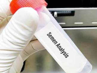
Figure 1
|
Figure 1 Semen
Collection |
3) CRIME
SCENE COLLECTION
· Surfaces: Semen can be collected from various surfaces at a crime scene, such as floors, furniture, or walls. Lee et al. (2001)
· Swabbing or Scraping: For non-porous surfaces, a moistened sterile swab is used. For porous surfaces, scraping may be necessary to collect the sample.
4) CONDOMS
· Used Condoms: If a used condom is found at a crime scene or provided by the victim, it can contain semen. The exterior and interior are swabbed to collect any biological material. Lehrman (1998)
· Handling: Careful handling and proper packaging are essential to avoid contamination.
5) UNDERWEAR
AND PERSONAL ITEMS
· Underwear: Victim’s or suspect’s underwear may contain semen stains and should be collected as evidence.
· Other Items: Personal items like tissues, sanitary products, or sex toys that may contain semen should also be collected.
4. BLOOD
Bloodstain pattern analysis (BPA) is a critical aspect of forensic science, helping investigators understand the events that led to the creation of bloodstains at a crime scene. Here’s an overview of how bloodstain patterns can be helpful in criminal investigations, the methods used, and some example cases:
4.1. BLOODSTAIN PATTERNS HELP IN INVESTIGATIONS [ NIST]
· Reconstructing Events: Bloodstain patterns can help reconstruct the events of a crime, indicating the positions and movements of the victim and assailant during the incident. Bevel & Gardner (2008)
· Determining Weapon Types: Different weapons create different bloodstain patterns. For example, a blunt object might produce different spatter patterns compared to a gunshot. Virkler & Lednev (2009)
· Identifying Struggle: The presence of cast-off patterns or void patterns can indicate a struggle or multiple blows. Bevel & Gardner (2008)
· Estimating Time of Events: The drying times and coagulation of blood can help estimate the time of events. James et al. (2005), Barrera et al. (2018)
· Linking Suspects: Blood patterns on suspects’ clothing can link them to the crime scene.
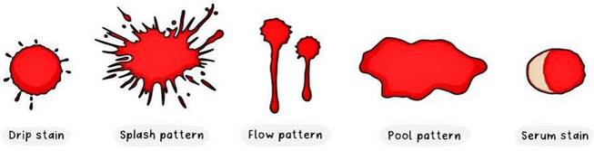
Figure 2
|
Figure 2 Different
Types of Blood Patterns 06 |
4.2. Methods of Bloodstain Pattern Analysis
· Visual Analysis: Initial examination involves visual inspection of the scene and documenting the bloodstains through photographs and sketches. Van Dam et al. (2019), Bevel & Gardner (2008)
4.3. TYPES OF PATTERNS
· Passive Stains: Result from the force of gravity (e.g., drops, pools, flows).
· Transfer Stains: Created when a bloody surface comes into contact with another surface.
· Projected Stains: Result from blood being subjected to an external force, such as a gunshot or blunt force.
· Angle of Impact: Analysts measure the shape of bloodstains to determine the angle at which the blood impacted a surface.
· Area of Origin: Using stringing or mathematical methods (e.g., trigonometry), analysts can determine the point where the blood originated.
· Velocity Analysis: Patterns are categorized based on the velocity of the blood source (low, medium, or high velocity).
1) EXAMPLE
CASES
The Jeffrey MacDonald Case (1970): Bloodstain
patterns were significant in the investigation of the murders of MacDonald’s
wife and daughters. The analysis of blood spatter, among other evidence, helped
convict MacDonald. Haglund
& Sorg (1997), Bevel
& Gardner (2008)
The David Camm Case (2000): Bloodstain analysis was pivotal in this case, where David Camm was accused of murdering his family. The interpretation of blood spatter patterns on his shirt was a key piece of evidence, although this case went through multiple trials and appeals. Haglund & Sorg (1997), Byrd & Castner (2001)
4.4. PRINCIPAL OF BLOOD GROUP IDENTIFICATION
Human red blood cells possessing A, B, and D antigens will agglutinate in the presence of antibodies directed toward the antigen. Agglutination of red blood cells with Anti-A, Anti-B, and Anti-D (IgM) reagents is a positive test result and indicates the presence of the corresponding antigen. The absence of agglutination of red blood cells with Anti-A, Anti-B, and Anti-D (IgM) reagents is a negative test result and indicates the absence of the corresponding antigen. Haglund & Sorg (1997)
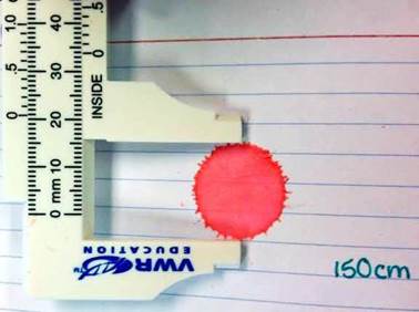
Figure 3
|
Figure 3 Measurement of
Dried Blood Stain |
1) PRELIMINARY
TEST FOR DRIED BLOOD GROUP
The middle finger was pricked, and blood from the veins in EDTA tubes the blood was spread across the tile surface. The blood was then allowed to air dry at a temperature of 23. 5 degrees Celsius for three hours. Each blood sample was collected using a spatula, and the dried blood sample was stored in various Eppendorf tubes that held five microliters of normal saline. Combination of regular saline in Eppendorf vials. Lee et al. (2020), Haglund & Sorg (1997), Byrd & Castner (2001)
2) TEST
PROCEDURE
· Place on drop each of the Anti-A, Anti-B, and Anti-D(RHO) (IgM) reagents on a clean glass slide.
· To each reagent drop, add one small drop (50 l) of whole blood.
· Rock the slide gently, back and forth.
· Observe for agglutination macroscopically at two minutes.
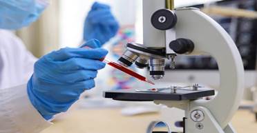
Figure 4
|
Figure 4 Microscopic
Examination |
Figure 5
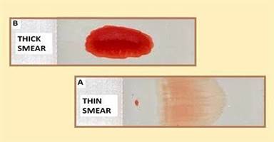
|
Figure 5
Thick-Smeared Slide, Thin-Stained Slide |
5. HAIR
Hair can be a valuable piece of evidence in crime investigations for several reasons:
5.1. DNA ANALYSIS
· Nuclear DNA: Hair follicles contain nuclear DNA, which can be used to identify individuals with high precision through DNA profiling.
· Mitochondrial DNA: Even if the hair lacks a root, mitochondrial DNA from the hair shaft can provide maternal lineage information, which can be useful in identifying individuals when nuclear DNA is unavailable. Haglund & Sorg (1997), Virkler & Lednev (2009)
5.2. MICROSCOPIC COMPARISON
· Structure: The microscopic structure of hair (cuticle, cortex, and medulla) can provide information about the hair’s origin, color, and any artificial treatment.
· Comparative Analysis: Microscopic comparison of hair found at a crime scene with that of a suspect can help establish a connection, though it is less definitive than DNA analysis.
5.3. CHEMICAL ANALYSIS
· Toxicology: Hair can absorb and retain substances, such as drugs, toxins, or poisons, providing a timeline of substance use.
· Elemental Analysis: Trace elements in hair can sometimes indicate environmental exposure, diet, or geographical location, contributing to profiling.
Figure 6
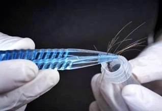
|
Figure 6 Collection of
Hair |
5.4. Growth Patterns
· Timeline: The growth rate of hair can help establish a timeline of events. For example, hair grows approximately 1 cm per month, so segments of hair can be analyzed to determine the timing of drug use or exposure to toxins. MacDonell (1971)
· Anomalies: Changes in hair growth patterns or damage can indicate trauma, stress, or other events relevant to the investigation.
5.5. ENVIRONMENTAL EVIDENCE
· Trace Evidence: Hair can pick up trace elements from the environment, such as pollen, dust, or soil, which can help investigators place a suspect at a particular location.
5.6. LINKING INDIVIDUALS
· Transfer Evidence: Hair can be transferred from one person to another during physical contact, helping establish connections between suspects, victims, and crime scenes.
5.7. SPECIES IDENTIFICATION
· Animal Hair: Sometimes, animal hair can be found at a crime scene, which can provide clues about the presence of pets or animals, leading to additional leads.
5.8. FORENSIC DATABASES
· Reference Collections: Forensic databases and reference collections of hair samples can aid in the comparison and identification process.
Hair evidence, when combined with other forensic techniques, can significantly contribute to solving crimes by providing valuable biological, chemical, and environmental information.
6. SALIVA
Saliva is a valuable source of forensic evidence in criminal investigations due to its biological properties and the information it can reveal. Here is a comprehensive overview of how saliva can be used in crime investigations:
6.1. IMPORTANCE OF SALIVA IN FORENSIC INVESTIGATIONS
· DNA Profiling: Saliva contains epithelial cells that carry DNA, which can be used to create a genetic profile of a suspect or victim. Lee et al. (2001)
· Biochemical Markers: Saliva contains enzymes, antibodies, and hormones that can provide additional information about a person’s health or drug use.
· Source Identification: Saliva can link a suspect to a crime scene or an object, such as a cigarette butt, glass, or bite mark.
6.2. COLLECTION METHODS
· Swabbing: Using sterile swabs to collect saliva from surfaces like the rim of a glass, cigarette butts, or bite marks.
· Absorption: Using absorbent materials to collect larger quantities of saliva.
· Cutting: Collecting saliva by cutting sections of fabric or other materials stained with saliva.
· Liquid Collection: Collecting liquid saliva directly from a person’s mouth using a collection device.
6.3. PRESERVATION OF SALIVA SAMPLES
· Dry Storage: Allowing saliva on swabs or other materials to air dry before storing them in paper envelopes or boxes. James et al. (2005)
· Refrigeration: Storing saliva samples in a refrigerator to prevent degradation.
· Use of Preservatives: Using chemical preservatives to stabilize saliva samples for long- term storage.
6.4. ANALYSIS METHODS
6.4.1. DNA EXTRACTION AND PROFILING
· Extraction: Isolating DNA from the cells present in the saliva.
· PCR (Polymerase Chain Reaction): Amplifying the DNA to generate enough material for analysis.
· STR (Short Tandem Repeat) Analysis: Creating a DNA profile by examining specific regions of the DNA. Lehrman (1998)
· Enzyme-Linked Immunosorbent Assay (ELISA): Detecting the presence of specific proteins or antibodies in saliva, which can indicate drug use or exposure to pathogens.
· Mass Spectrometry: Identifying and quantifying compounds present in saliva, useful for detecting drugs or poisons.
6.4.2. APPLICATIONS IN CRIME INVESTIGATIONS
· Linking Suspects to Crime Scenes: Saliva found on items at a crime scene can be matched to a suspect’s DNA, establishing their presence. James et al. (2005)
· Sexual Assault Cases: Saliva on the victim’s body or clothing can provide evidence of the assailant.
· Burglary and Theft: Saliva on items like glass shards, masks, or clothing can identify perpetrators.
· Homicide: Saliva on bite marks or other surfaces can link a suspect to the crime.
· Drug Use Detection: Saliva analysis can reveal recent drug use by suspects or victims, which can be relevant to the investigation.
6.5. EXAMPLE CASES
· The Amanda Knox Case: Saliva and DNA evidence were critical in the investigation of the murder of Meredith Kercher. Saliva on the victim’s body helped identify potential suspects.
· The Golden State Killer: In this case, DNA evidence from saliva was used decades after the crimes to identify and convict Joseph James DeAngelo.
· The Boston Strangler: DNA from saliva collected from a preserved evidence sample was used to confirm the identity of the perpetrator, Albert DeSalvo, many years after the murders.
7. FECAL MATERIAL
Fecal material can provide valuable forensic evidence in criminal investigations. Here are several aspects of how fecal material can be used in forensic science:
7.1. IDENTIFICATION OF INDIVIDUALS
·
DNA Analysis: Fecal matter contains cells
from the gastrointestinal tract, which can be used for DNA profiling. This
helps in identifying individuals if their DNA profile is already in the
database or for comparing with suspects. Gray (1977), Virkler
& Lednev (2009)
· Microbiome Analysis: The unique composition of gut bacteria (microbiome) can also be analyzed to identify individuals or determine lifestyle factors such as diet or geographic location. Gray (1977), Bevel & Gardner (2008) , Virkler & Lednev (2009)
7.2. DETERMINING TIME OF DEATH OR CRIME
· Bowel Content Examination: The state of digestion of the contents in fecal matter can help estimate the time of the last meal, which can be correlated with the time of death or the crime. James et al. (2005), Bevel & Gardner (2008)
7.3. INKING SUSPECTS TO CRIME SCENES
· Presence at the Scene: If fecal matter is found at a crime scene, it can link a suspect to that location.
· Transfer Evidence: Traces of fecal matter on clothing, footwear, or objects can be used to establish a connection between the suspect and the crime scene. Haglund & Sorg (1997)
7.4. ENVIRONMENTAL AND SITUATIONAL EVIDENCE
· Diet and Health Analysis: The composition of fecal matter can provide clues about the diet, medication, and health status of an individual, offering additional context to the investigation. Van Dam et al. (2019)
· Toxicology: Analysis of fecal matter can reveal the presence of drugs, toxins, or poisons, which can be relevant in cases of drug abuse or poisoning.
7.5. INVESTIGATIVE TECHNIQUES
· Chemical Analysis: Techniques such as Gas Chromatography-Mass Spectrometry (GC- MS) can identify specific chemical compounds present in fecal matter, aiding in toxicological investigations. James et al. (2005)
· Microscopic Examination: Microscopic analysis can reveal undigested food particles, parasites, or other biological materials that can provide additional information.
7.6. CHALLENGES AND CONSIDERATIONS
· Degradation: Fecal matter can degrade quickly, especially in adverse environmental conditions, which can affect the reliability of the evidence.
· Contamination: Care must be taken to avoid contamination of fecal samples during collection and analysis to ensure the integrity of the evidence.
Fecal material, while not as commonly used as blood or saliva, can be a critical piece of evidence in certain forensic investigations. Advances in DNA technology and microbiome analysis are enhancing the potential applications of fecal evidence in forensic science.
8. URINE
The collection of urine can be done in various settings, including clinical, forensic, and research environments. The methodology for urine collection can vary depending on the purpose of the analysis.
8.1. URINE COLLECTION METHODS
8.1.1. RANDOM URINE SAMPLE
· Procedure: The individual urinates into a clean container at any time of the day without any prior preparation.
· Uses: Suitable for routine tests and initial screenings.
8.1.2. FIRST MORNING URINE SAMPLE
· Procedure: The first urine passed after waking up is collected.
· Uses: Ideal for detecting substances that may be more concentrated in the urine after an overnight fast, such as hCG in pregnancy tests.
8.1.3. MIDSTREAM CLEAN-CATCH SAMPLE
· Procedure: The individual cleans the genital area before urinating and collects urine midstream in a sterile container to minimize contamination. James et al. (2005)
· Uses: Commonly used for bacterial cultures and infection detection. Timed Urine Collection:
· Procedure: Urine is collected over a specified period, such as 24 hours, to measure the excretion of certain substances.
· Uses: Essential for quantitative tests like creatinine clearance, protein, or hormone levels.
8.1.4. CATHETERIZED URINE SAMPLE
Figure 7
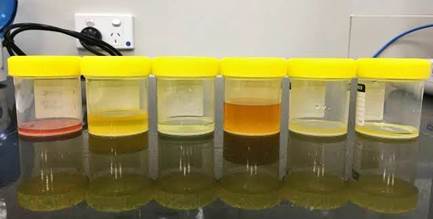
|
Figure 7 Different
Properties of Urine |
· Procedure: A sterile catheter is inserted through the urethra into the bladder to obtain a urine sample.
· Uses: Used when a clean-catch sample is not possible, such as in individuals with mobility issues or severe illness. Lee et al. (2001)
8.1.5. SUPRAPUBIC ASPIRATION
· Procedure: A needle is inserted through the abdominal wall into the bladder to collect urine.
· Uses: Used primarily in infants or when contamination needs to be completely avoided.
8.2. METHODOLOGY FOR URINE ANALYSIS
8.2.1. PHYSICAL EXAMINATION
· Color: Urine color can indicate hydration status and the presence of certain substances.
· Clarity: Clear, cloudy, or turbid urine can indicate different conditions.
· Odor: Unusual odors can be a sign of metabolic or infectious diseases.
8.2.2. CHEMICAL ANALYSIS
· Dipstick Tests: Quick tests using a reagent strip to detect pH, specific gravity, protein, glucose, ketones, bilirubin, urobilinogen, nitrite, and leukocyte esterase.
· Spectrophotometry: Used for more precise measurements of substances like glucose, creatinine, or electrolytes.
8.2.3. MICROSCOPIC EXAMINATION
· Sediment Analysis: Urine is centrifuged, and the sediment is examined under a microscope for cells, crystals, bacteria, and other particles.
8.2.4. MICROBIOLOGICAL ANALYSIS
· Culture and Sensitivity: Urine is cultured to detect and identify bacteria, fungi, or other pathogens, and to determine antibiotic susceptibility. James et al. (2005)
8.2.5. ADVANCED TECHNIQUES
· Mass Spectrometry: For detailed analysis of small molecules and metabolites.
· Polymerase Chain Reaction (PCR): For detecting genetic material from pathogens.
8.3. CONSIDERATIONS AND BEST PRACTICES
· Sterility: Ensure that collection containers and methods are sterile to prevent contamination.
· Labeling: Properly label all samples with the patient’s information, date, and time of collection.
· Storage: Store samples at appropriate temperatures to preserve integrity, especially for timed collections.
· Documentation: Record any medications, dietary intake, or activities that may influence urine composition.
By following these methodologies and best practices, urine collection and analysis can provide critical insights for diagnostic, forensic, and research purposes. James & Nordby (2009), Gray (1977), Byrd & Castner (2001)
9. SKIN TISSUE
Skin collection in forensic investigations involves obtaining samples of skin or skin cells from a crime scene or a person to help establish a connection, identify individuals, or gather other crucial evidence.
9.1. METHODOLOGY FOR SKIN COLLECTION
9.1.1. SURFACE SWABBING
· Procedure: Use sterile cotton swabs moistened with sterile water or saline to swab the skin surface where evidence is suspected.
· Use: Collects skin cells, DNA, and other biological material from surfaces or objects that may have come into contact with a person.
9.1.2. TAPE LIFTING
Figure 8
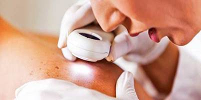
|
Figure 8 Skin
Visualization |
· Procedure: Use adhesive tape to lift skin cells and other particulate matter from surfaces.
· Use: Effective for collecting trace evidence, such as skin cells, hair, fibers, and other minute particles.
9.1.3. SCRAPING
· Procedure: Gently scrape the skin or a surface with a sterile instrument to collect cells and biological material.
· Use: Used when a more concentrated sample is needed, often from surfaces with visible biological material.
9.2. COLLECTION OF SKIN TISSUE
· Procedure: In cases of injury or post-mortem examination, small samples of skin tissue may be collected using sterile surgical instruments. James & Eckert (1999), Bevel & Gardner (2008), Virkler & Lednev (2009)
· Use: Provides DNA and histological analysis; useful in cases of violent crime where skin may be under fingernails or on weapons.
9.3. VACUUM COLLECTION
· Procedure: Use a forensic vacuum device with a filter to collect skin cells and other particulate matter from larger areas.
· Use: Useful for collecting trace evidence from carpets, furniture, and large surfaces.
9.4. FORENSIC AUTOPSY
· Procedure: During an autopsy, skin samples are collected using scalpels or biopsy punches.
· Use: Provides detailed information about injuries, disease, and identification in death investigations.
9.5. ANALYSIS OF SKIN SAMPLES
9.5.1. DNA PROFILING
· Procedure: Extract DNA from collected skin cells and analyze using Polymerase Chain Reaction (PCR) or Short Tandem Repeat (STR) analysis. James & Nordby (2009), James et al. (2005), Virkler & Lednev (2009)
· Use: Identifies individuals, links suspects to crime scenes, or excludes individuals from suspicion.
9.5.2. MICROSCOPIC EXAMINATION
· Procedure: Examine skin cells and tissue under a microscope to identify cellular structures, injuries, or foreign particles.
· Use: Provides insights into the nature of injuries, presence of foreign materials, or signs of disease.
9.5.3. CHEMICAL ANALYSIS
· Procedure: Analyze chemical residues on the skin using techniques such as Gas Chromatography-Mass Spectrometry (GC-MS).
· Use: Identifies substances such as drugs, toxins, or environmental contaminants.
9.5.4. HISTOLOGICAL EXAMINATION
· Procedure: Prepare and stain skin tissue sections for examination under a microscope.
· Use: Reveals details about skin structure, pathology, and trauma.
9.6. CONSIDERATIONS FOR FORENSIC SKIN COLLECTION
9.6.1. CONTAMINATION PREVENTION
· Procedure: Use sterile tools and techniques to prevent contamination of samples.
· Use: Ensures the integrity and reliability of collected evidence. Laber & Epstein (1983)
9.6.2. CHAIN OF CUSTODY
· Procedure: Document every step of the collection, storage, and analysis process.
· Use: Maintains legal and evidentiary standards, ensuring that evidence is admissible in court.
9.6.3. PROPER STORAGE
· Procedure: Store collected samples in appropriate conditions, such as refrigeration or freezing, depending on the analysis required.
· Use: Preserves the sample’s integrity for accurate analysis.
9.6.4. ETHICAL AND LEGAL CONSIDERATIONS
· Procedure: Obtain proper consent and follow legal protocols, especially in living subjects.
· Use: Ensures compliance with ethical standards and legal requirements. Laber & Epstein (1983), Bevel & Gardner (2008), Virkler & Lednev (2009)
10. OBSERVATION
The search of evidence at the crime scene is observed by bloodstain pattern analyst for proper investigation at the scene. The retrieval of bloodstain pattern evidence begins with the effective search of the scene. Physical evidence refers to any tangible article, small or large, which tends to prove or disprove a point in question. Consider the murder held in India in an area where security is considerably high.
11. RESULT
Violent criminal acts are often accompanied by dynamic blood shedding events at the crime scene. Bloodstain pattern analyst particularly deals with estimation of the dynamic blood shedding events from the static bloodstain patterns that have been left at the scene. Of all the stain patterns present at a crime scene, drip stain patterns are common stain patterns one would expect to document at a violent crime scene. The paper describes the procedural steps that are taken by the bloodstain pattern analyst during the investigation of a crime.
12. ABBREVIATION
|
A – SHORT |
B- FULL |
|
AAFS |
American Academy of Forensic Sciences |
|
ANSI |
American National Standards Institute |
|
ASB |
AAFS Standards Board |
|
BPA |
Bloodstain Pattern Analysis |
|
DNA |
D-deoxyribonucleic acid |
|
FSR |
Forensic Science Regulator |
|
HVIS |
High-Velocity Impact
Spatter |
|
IABPA |
International Association of Bloodstain
Pattern Analysts |
|
ISO |
International Organization for Standardization |
|
MVIS |
Medium Velocity Impact |
|
NIST |
National Institute of Standards and Technology |
|
OSAC |
Organization
of Scientific Area
Committees |
|
PT |
Proficiency testing |
|
PCR |
Polymerase Chain Reaction |
|
STR |
Short Tandem Repeat |
|
SWGSTAIN |
Scientific Working Group for Blood Group Pattern Analysis |
|
WCC |
Wildlife Conservation Centre |
|
WGRB |
Wildlife Genomic Resource Bank |
|
ZF |
Zinc Finger |
13. CONCLUSION
Biological evidence plays a crucial role in crime investigation by providing objective and scientifically verifiable data that can significantly influence the outcome of criminal cases. This type of evidence, which includes DNA, blood, hair, skin cells, and other bodily substances, offers a unique means of linking suspects to crime scenes, victims, or criminal activities. Advances in forensic science have enhanced the accuracy and reliability of analyzing biological materials, making it possible to identify individuals with a high degree of certainty, even in cases where other forms of evidence might be lacking. The utilization of DNA profiling, In particular, has revolutionized forensic science by enabling law enforcement agencies to solve cold cases, exonerate the wrongly accused, and establish connections between seemingly unrelated crimes. This has been made possible through the development of sophisticated techniques such as polymerase chain reaction (PCR) and short tandem repeat (STR) analysis, which allow for the examination of even the smallest biological samples. However, the interpretation and handling of biological evidence require meticulous care to prevent contamination, degradation, or misinterpretation, which could lead to wrongful convictions or the failure to identify the true perpetrator. Proper chain-of- custody protocols, rigorous laboratory procedures, and the use of accredited forensic experts are essential to ensuring the integrity of biological evidence throughout the investigative process. In conclusion, biological evidence serves as a powerful tool in crime investigation, offering the potential for definitive identification of individuals involved in criminal activities. While it is not infallible, when handled and interpreted correctly, it provides a strong foundation for achieving justice. As forensic technology continues to evolve, the role of biological evidence in crime investigation is likely to expand, further enhancing the ability of law enforcement to solve crimes and uphold the rule of law.
CONFLICT OF INTERESTS
None.
ACKNOWLEDGMENTS
The government has supported me to publish this review article. Thank you so much. It has been written by me myself. I am currently pursuing master’s [2024]. Thank you National Crime Bureau of Investigation [NCBI] Government of India.
REFERENCES
Barrera, V., Haas, C., Meixner, E.A., & Fliss, B. (2018). Detection of Painted-Over Traces of Blood and Seminal Fluid. Int J Legal Med, 132(4), 1067-1074. https://doi.org/10.1007/s00414-018-1787-7
Bevel, T., & Gardner, R. M. (2008). Bloodstain Pattern Analysis: With an Introduction to Crime Scene Reconstruction, (3rd Ed.). Boca Raton, FL: CRC Press.
Bonaccordo, E. (2018). Evaluation of an Automated Panoramic Imaging System for the Photographic Recording and Analysis of Blood Spatter in Crime Scenes. (M. Res.) Western Sydney University (Australia) Ann Arbor. ProQuest Dissertations & Theses A&I Database.
Boonkhong, K., Karnjanadecha, M., & Aiyarak, P. (2010). Impact Angle Analysis of Bloodstains Qusing a Simple Image Processing Technique Songklanakarin. J. Sci. Technol, 32(2), 169-173.
Byrd, J. H., & Castner, J. L. (2001). Forensic Entomology: The Utility of Arthropods in Forensic Investigations. Boca Raton, FL: CRC Press.
Cotton, R. W., & Fisher, M. B. (2015). Review: Properties of Sperm and Seminal Fluid, Informed by Research on Reproduction and Contraception. Forensic Science International. Genetics, 18, 66–77. https://doi.org/10.1016/j.fsigen.2015.03.009
DeForest, P. R., Gaensslen, R. E., & Lee, H. C. (1983). Forensic Science: An Introduction to Criminalistics. New York: McGraw-Hill.
DiMaio, V. J. M. (1999). Gunshot Wounds: Practical Aspects of Firearms, Ballistics and Forensic Techniques, (2nd Ed). Boca Raton, FL: CRC Press.
Gardner, R. M. (2004). Practical Crime Scene Processing and Investigation. Boca Raton, FL: CRC Press.
Gray, H. (1977). Gray’s Anatomy. New York: Crown Publishers.
Haglund, W. D., & Sorg, M. H. (1997). Forensic Taphonomy: The Postmortem Fate of Human Remains. Boca Raton, FL: CRC Press.
James, S. H. (1999). Scientific and Legal Applications of Bloodstain Pattern Interpretation. Boca Raton, FL: CRC Press.
James, S. H., & Eckert, W. G. (1999). Interpretation of Bloodstain Evidence at Crime Scenes, (2nd Ed). Boca Raton, FL: CRC Press.
James, S. H., & Edel, C. F. (1997). “Bloodstain Pattern Interpretation,” in Introduction to Forensic Sciences, W. E. Eckert, Ed. Boca Raton, FL: CRC Press.
James, S. H., & Nordby, J. J. (2009). Forensic Science: An Introduction to Scientific and Investigative Techniques, (3rd Ed). Boca Raton, FL: CRC Press.
James, S. H., Kish, P. E., & Sutton, T. P. (2005). Principles of Bloodstain Pattern Analysis: Theory and Practice. Boca Raton, FL: CRC Press.
James, S. H., Kish, P. E., & Sutton, T. P. (2005). “Recognition of Bloodstain Patterns, ” in Forensic Science: An Introduction to Scientific and Investigative Techniques, (2nd Ed.), S. H. James and J. J. Nordby, Eds. Boca Raton, FL: CRC Press.
Kirk, P. L. (1974). Crime Investigation, (2nd Ed). New York: John Wiley & Sons.
Laber, T. L., & Epstein, B. P. (1983). Bloodstain Pattern Analysis. Minneapolis, MN: Callen Publishing.
Lee, H. C., Palmbach, T. M., & Miller, M. T. (2001). Henry Lee’s Crime Scene Handbook. San Diego, CA: Academic Press.
Lee, S. Y., Seo, Y. I., Moon, B. S., Kim, J. P., Goh, J. M., Park, N. K., & Shin, S. H. (2020). Study on Development of Forensic Blood Substitute: Focusing on Bloodstain Pattern Analysis. Forensic Science International, 316. https://doi.org/10.1016/j.forsciint.2020.110461
Lehrman, R. L. (1998). Physics: The Easy Way, (3rd Ed). Hauppaugh, New York.
Lu, X., Xu, Z., Niu, Q.S., & Tu, Z. (2018). Application of Touch DNA in Investigation Practice. Fa Yi Xue Za Zhi, 34(3), 294-298. https://doi.org/10.12116/j.issn.1004-5619.2018.03.015
MacDonell, H. L. (1971). “Interpretation of Bloodstains: Physical Considerations, ” in Legal
Medicine Annual, C. Wecht, Ed. New York: Appleton, Century Crofts.
MacDonell, H. L. (1981). “Criminalistics, Bloodstain Examination, ” in Forensic Sciences, Vol.
3, C. Wecht, Ed. New York: Matthew Bender.
MacDonell, H. L. (2005). Bloodstain Patterns, (2nd rev. Ed). Corning, NY: Laboratory of Forensic Science.
McMichael, G. L., Gibson, C. S., O'Callaghan, M. E., Goldwater, P. N., Dekker, G. A., Haan, E. A., MacLennan, A. H., & South Australian Cerebral Palsy Research Group (2009). DNA from Buccal Swabs Suitable for High-Throughput SNP Multiplex Analysis. Journal of Biomolecular Techniques: JBT, 20(5), 232–235.
Thanakiatkrai, P., Raham, K., Pradutkanchana, J., Sotthibandhu, S., & Kitpipit, T. (2017). Direct-STR Typing from Presumptively-Tested and Untreated Body Fluids. Forensic Sci Int Genet, 30, 1-9. https://doi.org/10.1016/j.fsigen.2017.06.001
Van Dam, A., Schoon, A., Wierda, S. F., Heeringa, E., & Aalders, C. G. (2019). The Use of Crime Scene Detection Dogs to Locate Semen Stains on Different Types of Fabric. Forensic Science International, 302.
Virkler, K., & Lednev, I.K. (2009). Analysis of Body Fluids for Forensic Purposes: from Laboratory Testing to Non- Destructive Rapid Confirmatory Identification at a Crime Scene. Forensic Sci Int, 188(1-3), 1-17. https://doi.org/10.1016/j.forsciint.2009.02.013
|
|
 This work is licensed under a: Creative Commons Attribution 4.0 International License
© DigiSecForensics 2024. All Rights Reserved.